


IV-HSL Emitter Cell
Abstract
The Emitter Cell is a synthetic cell that produces and releases the signaling molecule IV-HSL to communicate with E. coli bacteria, serving as a foundation for creating Responder Cells that can detect molecular inputs and amplify signals in co-culture systems.
Overview
The Emitter Cell produces and releases a chemical signal molecule into the environment. This capacity provides an example of enzymatic small-molecule production, molecule release as a reporter output, inter-cell communication, and co-culture of synthetic cells with living bacteria. The Emitter Cell is largely based on a paper by Jefferson M Smith, Denis Hartmann, and Michael J. Booth: Engineering cellular communication between light-activated synthetic cells and bacteria.
In this first Emitter, the cell produces and releases N-isovaleryl-L-homoserine lactone (IV-HSL). IV-HSL is a branched acyl-homoserine lactone with several advantages for use in the Emitter Cell: it is able to cross the synthetic cell membrane; its uncommon branched-chain structure makes it orthogonal from many other HSLs Lindemann, 2011; and it is able to activate expression in signal receiver cells (in this case, E. coli) at very low (picomolar) concentrations Lindemann, 2011. The IV-HSL signal is received by a population of E. coli cells, which respond by producing a fluorescent output.
The Detector Cells and Emitter together form the basis for an upcoming Responder Cell; a synthetic cell which can detect a molecular input (such as aTc or IV-HSL itself), and produce a molecular output (IV-HSL) in response. Coupling Detector and Emitter modules will enable signal amplification, where a low amount of a molecule of interest can activate a large population of Responder cells and generate an output that is easy to detect. IV-HSL-detecting Responder Cells could also detect the production of IV-HSL from living cells, providing a means to report on their state in co-culture.

Figure 1:Schematic of IV-HSL emitter cell.
Design
The Emitter Cell implements the IV-HSL Emitter Module within a synthetic cell. The Emitter Module produces the BjaI enzyme under the control of a constitutive T7 promoter. BjaI produces IV-HSL from two substrate molecules, S-adenosylmethionine (SAM) and isovaleryl coenzyme A (IV-CoA). IV-HSL diffuses out of the cell, through the lipid bilayer.

Figure 2:Schematic of IV-HSL emitter module.
Usage
Protocol
There are five key stages to making the IV-HSL Emitter Cell:
| Step | Process | Hands-on Time | Total Time | Notes |
|---|---|---|---|---|
| 1 | Pre-culture BjaR receiver cells | 30 mins | 3.5 hr | |
| 2 | Prepare lipids-in-oil solution, outer solution, and substrate stock solutions | 1 hr | 4 h | Buffers and lipids may be prepared in advance and used for experiments on subsequent days. |
| 3 | Assemble PURE reactions | 30 mins | 30 mins | |
| 4 | Encapsulate liposomes | 30 mins | 30 mins | |
| 5 | Measure and image | 30 mins | 6–12 h | Total time depends on the exact experiment and incubation conditions. GFP expression should be seen over the first 6 hours at 37C. |
Step 1: Pre-culture BjaR receiver cells
Prepare glycerol stock of BjaR receiver cells
Transform XL-10 Gold competent E. coli with
bjaR-GFP-native:Add 1–5 µl containing 1 pg–100 ng of plasmid DNA
bjaR-GFP-nativeto 50 µl of XL10-Gold cell mixture. Carefully flick the tube 4–5 times to mix cells and DNA. Do not vortex.Place the mixture on ice for 15 minutes. Do not mix.
Heat shock at exactly 42°C for 40 seconds. Do not mix.
Place on ice for 5 minutes. Do not mix.
Pipette 950 µl of room temperature SOC into the cell mixture.
Shake the cell mixture vigorously (250 rpm) at 37°C for 60 minutes.
Warm Ampicilin LB agarose plates at 37°C for 10 mins.
Mix the cells thoroughly by flicking the tube and inverting, then perform several 10-fold serial dilutions in LB.
Spread 50–100 µl of each dilution onto a Ampicilin agarose plate and incubate overnight for ~15 hrs at 37°C.
Prepare a streak plate from the glycerol stock (reference)
Streak a Ampicillin LB plate from the glycerol stock and incubate overnight at 37C.
Prepare M9 Media containing 1× M9 salts, 0.34 mg/ml−1 thiamine hydrochloride, 0.2% casamino acids, 2 mM MgSO4, 100 µM CaCl2 and 0.4% (wt/vol) glucose.
Pick a colony from the E. coli streak plate, and inoculate a 5 mL culture tube containing the M9 media with 100 ug/mL carbenicillin.
Incubate the cells at 37 °C, 225 rpm, for 3 h. Prepare Emitter liposomes while the cells incubate.
Dilute the culture media with the pre-warmed M9 media until OD600 = ~0.1.
Balance osmolarity of the culture media with PURE (inner solution in liposomes) by adding glucose to the M9 media:
| Volume to mix (uL) | |
|---|---|
| M9 media | 1000 |
| 3M Glucose | 293.81 |
Step 2: Prepare lipids-in-oil solution, outer solution, and substrate stock solutions
Prepare lipids-in-oil (mineral oil) solution.
Clean glass syringes. Pour a small amount of 95% ethanol into a glass container (e.g. a 10 mL beaker).Assemble the glass syringe and prime it by drawing ethanol into the glass syringe, then empty into a waste bottle.
Use glass syringes to add lipids, as shown in the table below, into the 10 ml glass vial containing 1 ml of mineral oil (final lipid concentration is 5 mg/ml).
Heat the lipids-in-oil mixture on a hotplate at 55 C for 3 hrs.
Vortex the lipids-in-oil mixture for 1 min.
The lipids-in-oil mixture can be stored at 4 C for up to 3 days.
| Lipids | Stock Concentration (mg/mL) | Volume to add (uL) | Target percentage |
|---|---|---|---|
| Egg PC | 25 | 160 | 66.68 |
| Cholesterol | 50 | 20 | 33.32 |
| 18:0 Liss Rhod PE | 1 | 5 | 0.01 |
Prepare outer solution. Final concentration of sugar stock solution is 900 mM
| Buffer | Volume to add (uL) |
|---|---|
| 3M Glucose Stock | 700 |
| H2O | 300 |
Prepare substrate stock solutions
| Substrate | Concentration (uM) | MW (g/mol) | Weight (g) | Final Volume (mL) |
|---|---|---|---|---|
| SAM | 5000 | 398.44 | 1.99 | 1 |
| IV-CoA | 5000 | 851.65 | 4.26 | 1 |
| IV-HSL | 10 | 183.21 | 1.83 | 1 |
Step 3: Assemble PURE Reactions
PURE reaction setup
| Sample | Negative control | Positive control | ||
|---|---|---|---|---|
| Component | Volume (uL) | Volume (uL) | Volume (uL) | Notes |
| PURE Solution A | 12 | 12 | 0 | PURE energy solution: small molecules |
| PURE Solution B | 9 | 9 | 0 | PURE proteins and ribosomes |
| RNAse Inhibitor | 1.5 | 1.5 | 0 | Prevents RNAse activity |
| EM01-pOpen-pT7-BjaI (~200 ng/uL) | 1.5 | 0 | 0 | DNA encoding green fluorescent protein |
| SAM (5mM) | 1.8 | 1.8 | 0 | Substrate for IV-HSL production. |
| IV-CoA (5mM) | 0.48 | 0.48 | 0 | Substrate for IV-HSL production. |
| OptiPrep | 1.5 | 1.5 | 1.5 | Adds density for phase-transfer |
| IV-HSL (10 uM) | 0 | 0 | 0.3 | Commercial IV-HSL for positive control. |
| 3M Glucose | 0 | 0 | 8.46 | |
| ddH2O | 2.22 | 3.72 | 19.74 | |
| Total | 30 | 30 | 30 |
Thaw reagents on ice and then keep on ice.
Prepare a PCR strip in a strip holder on ice for assembly of the three reactions (Sample, Negative, Positive).
Step 4: Encapsulate PURE reactions into Liposomes
Some tips and tricks can be found in PURE Cell.
Set up a microfuge tube rack, with three 1.5 mL microfuge tubes per liposome encapsulation:
Number the tubes per the number of reactions assembled in Step 3.
For each reaction, label the two tubes:
I — Oil emulsion
O — Outer solution
Add 30 ul of PURE reactions prepared in Step 3 to tubes labelled I.
Add 180 uL of the lipids-in-oil mixture on top of the PURE reactions in tubes labelled I and pipette vigorously until the emulsion becomes cloudy.
Add 300 uL of outer solution to each of the tubes labelled O.
Add 210 uL of the milky solution carefully on top of the outer solution in the tubes labelled O.
Centrifuge at 9000 rpm at 4c for 10 mins.
Remove the top oil and resuspend the pellet in 100 ul of outer solution.
Collect the liposomes.
Step 5: Measure and Image Liposomes and Cells
Imaging using confocal microscopy (Operetta CLS):
While microscopy setups may vary, our performance data was collected using the following configuration.
Add BjaR receiver cells prepared in Step 1 into 384 Well Glass Bottom Microplates.
Add 10 uL of liposomes made in Step 3 on top of the receiver cells in 384 Well Glass Bottom Microplates.
Imaging conditions using Operetta:
Temperature: 37 C degree
Green fluorescence channel (200 us expsoure 95%) - excitation: 460-490 nm; emission: 500-550 nm.
Red fluorescence channel (50 us exposure 95%) - excitation: 530-560 nm; emission: 570-650 nm.
Brightfield (20 us 95%)
We capture a 6 h time lapse with 10 min intervals.
We also acquired z-stack images spanning from 0 µm to 80 µm of the focal plane.
Measuring usinng plate reader (BioTek Cytation 5):
Add BjaR receiver cells prepared in Step 1 into 96 Well Glass Bottom Microplates.
Add 10 uL of liposomes made in Step 3 on top of the receiver cells in 96 Well Glass Bottom Microplates.
Procedures:
Temperature: 37 C degree
Read the fluorescence intensity from the bottom
Excitation wavelength: 485 nm ; Emission wavelength: 528 nm
We capture a 6 h time lapse with 5 min intervals
Modules
DNA Components
| Name | Length (bp) | Description | Link |
|---|---|---|---|
pT7-bjaI | 2752 | upcoming. Expresses the BjaI enzyme to produce IV-HSL. | pOpen-pT7-bjaI.gb |
bjaR-GFP-native | 3877 | upcoming. E. coli native receiver module; responds to IV-HSL by producing GFP. | pOpen |
Key Materials
| Name | Product | Manufacturer | Part # | Price | Link |
|---|---|---|---|---|---|
| Buffers | |||||
| Glucose | D-(+)-Glucose, 99% | Thermo Scientific | A16828-36 | $41.65 | [link] |
| Sucrose | Sucrose, bioultra, for molecular biology, ≥99.5% (HPLC) | Sigma-Aldrich | 84097-1KG | $170.00 | [link] |
| Lipids | |||||
| Egg PC | 25mg/mL | Avanti Lipids | 840051C-200mg | $186 | [link] |
| Cholesterol | Cholesterol (plant) | Avanti Research | A80100 | $261.00 | [link] |
| Liss-Rhod-PE | 18:0 Liss Rhod PE 1 mg/mL | Avanti Lipids | A81179 | $273.47 | [link] |
| Mineral Oil | Mineral oil, mixed weight | Thermo Scientific | AC415080010 | $53.40 | [link] |
| Glass Syringe 250 uL | Hamilton glass syringe | Hamilton | 14-815-238 | $150.15 | [link] |
| PURE | |||||
| PURE | PURExpress® In Vitro Protein Synthesis Kit | NEB | E6800L | $2774.00 | [link] |
| RNase Inhibitor | RNase Inhibitor, Murine | NEB | M0314S | $87.00 | [link] |
| DNA | pT7-bjaI | b. next | [link] | ||
bjaR-GFP-native | b.next | [link] | |||
| OptiPrep | OptiPrep™ | STEMCELL Technologies | 07820 | $289.00 | [link] |
| SAM | S-adenosylmethionine (SAM) | NEB | B9003S | $45 | [link] |
| IV-CoA | Isovaleryl coenzyme A lithium salt hydrate | Millipore Sigma | I9381-10MG | $348 | [link] |
| IV-HSL | 3-Methyl-N-[(3S)-tetrahydro-2-oxo-3-furanyl]butanamide | LGC | TRC-M282980-50MG | $171 | [link] |
| DMSO | Dimethyl sulfoxide | Thermo Scientific | 042780.M1 | $342 | [link] |
| Cell culture | |||||
| XL10-Gold Cells | XL10-Gold Ultracompetent Cells | Agilent | 200314 | $223 | [link] |
| M9 Media | M9, Minimal Salts, 5X, powder, minimal microbial growth medium | Sigma-Aldrich | M6030-1KG | $260 | [link] |
Performance Data
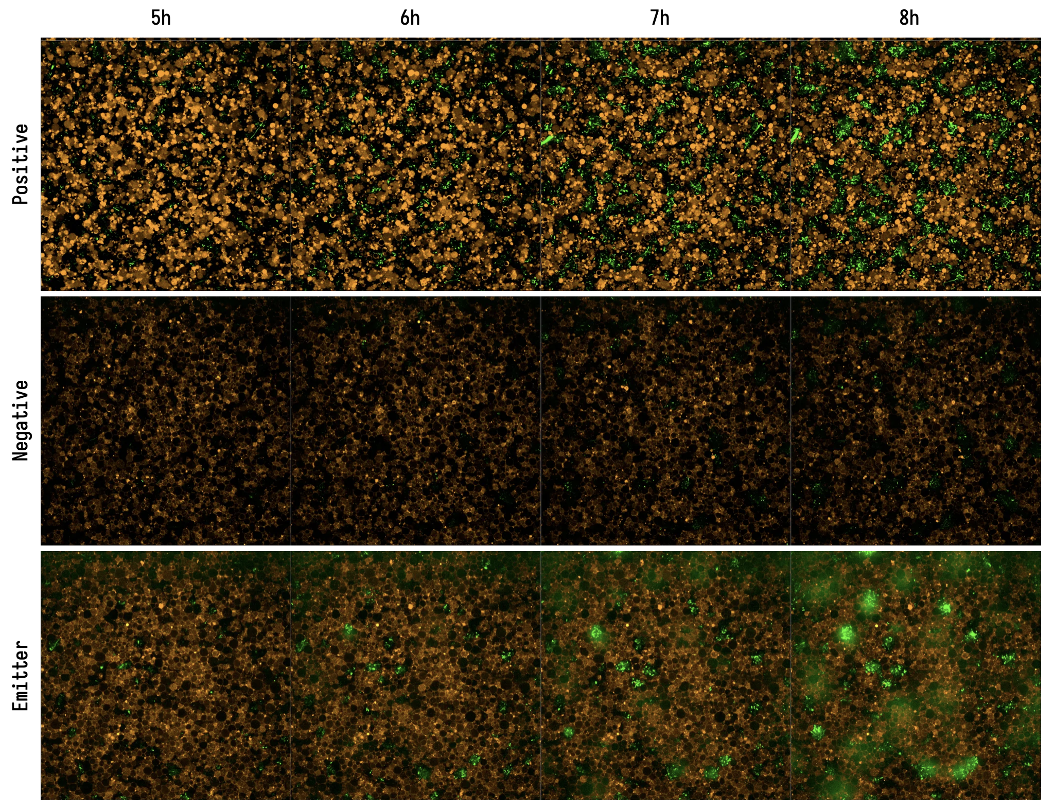
Emitter Cells were constructed following and co-cultured with E. coli containing the bjaR-GFP-native IV-HSL receiver plasmid. We performed time-series confocal microscopy (Revvity Operetta CLS) over 8 hours, collecting red (Rhodamine-B) and green (GFP) fluorescence, and brightfield images at 40x magnification across multiple fields per well, such that the entirety of each well was imaged. Timepoints were approximately 15 minutes apart.
The Emitter Cell causes E. coli to express GFP in response to IV-HSL.
Emitter Cell Timeseries. (Positive) Liposomes contain PURE and 100 nM IV-HSL. (Negative) Liposomes contain PURE supplemented with SAM and IV-HSL, but no DNA encoding BjaI. (Emitter) Liposomes contain PURE expressing BjaI from pT7-bjaI. Exposures are matched between wells. Each field of view is 167 uM wide.
Liposomes exclude E. coli cells from the plate coverslip
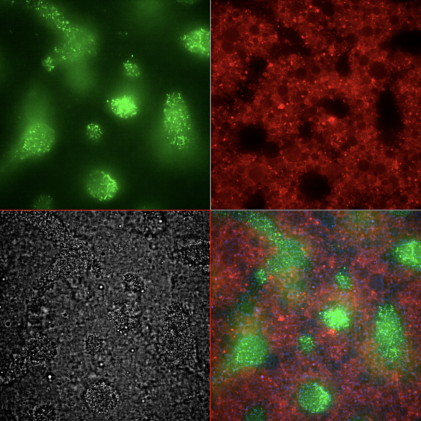
Figure 5:Emitter Cell Endpoint Montage. Single field of view of Emitter Cell in co-culture with E. coli receiver cells at t = 8 hours. (green) E. coli producing GFP in response to IV-HSL emitted by the emitter cells. (red) Emitter cells with rhodamine-labeled membrane producing IV-HSL. (grey) Brightfield image of liposomes and E. coli cells. (rgb) Merged image.
Figure 6:Emitter Cell Endpoint Z-Stack. Z-stack, single field of view of the Emitter Cell after the final timeseries timepoint (>8h). Liposomes preferentially form a layer on the surface of the cover slip, occluding many of the E. coli cells from the bottom layer imaged during the timeseries. More activated E. coli cells become visible at longer focal distances (higher in the liquid column of the well).
Background Protocols
Prepare lipids for use in encapsulation: Lipid Preparation
Prepare inner and outer buffers: PURE inner and outer solution
Credits
Jefferson Smith & Michael Booth (Oxford / UCL)
b.next
- Smith, J. M., Hartmann, D., & Booth, M. J. (2023). Engineering cellular communication between light-activated synthetic cells and bacteria. Nature Chemical Biology, 19(9), 1138–1146. 10.1038/s41589-023-01374-7
- Lindemann, A., Pessi, G., Schaefer, A. L., Mattmann, M. E., Christensen, Q. H., Kessler, A., Hennecke, H., Blackwell, H. E., Greenberg, E. P., & Harwood, C. S. (2011). Isovaleryl-homoserine lactone, an unusual branched-chain quorum-sensing signal from the soybean symbiont Bradyrhizobium japonicum. Proceedings of the National Academy of Sciences, 108(40), 16765–16770. 10.1073/pnas.1114125108